DNA only accounts for about half of the total volume of chromosomes, as shown by new 3D images of human chromosomes.
Until now, it has been generally accepted that chromosomes are mostly made up of DNA, with some proteins (histones) present to protect the DNA and prevent the chromosome from unravelling – this complex is known as chromatin. However, the small size of chromosomes has made detailed study of their structure difficult as they fall between the optimum sizes for imaging with a light microscope or an electron microscope.
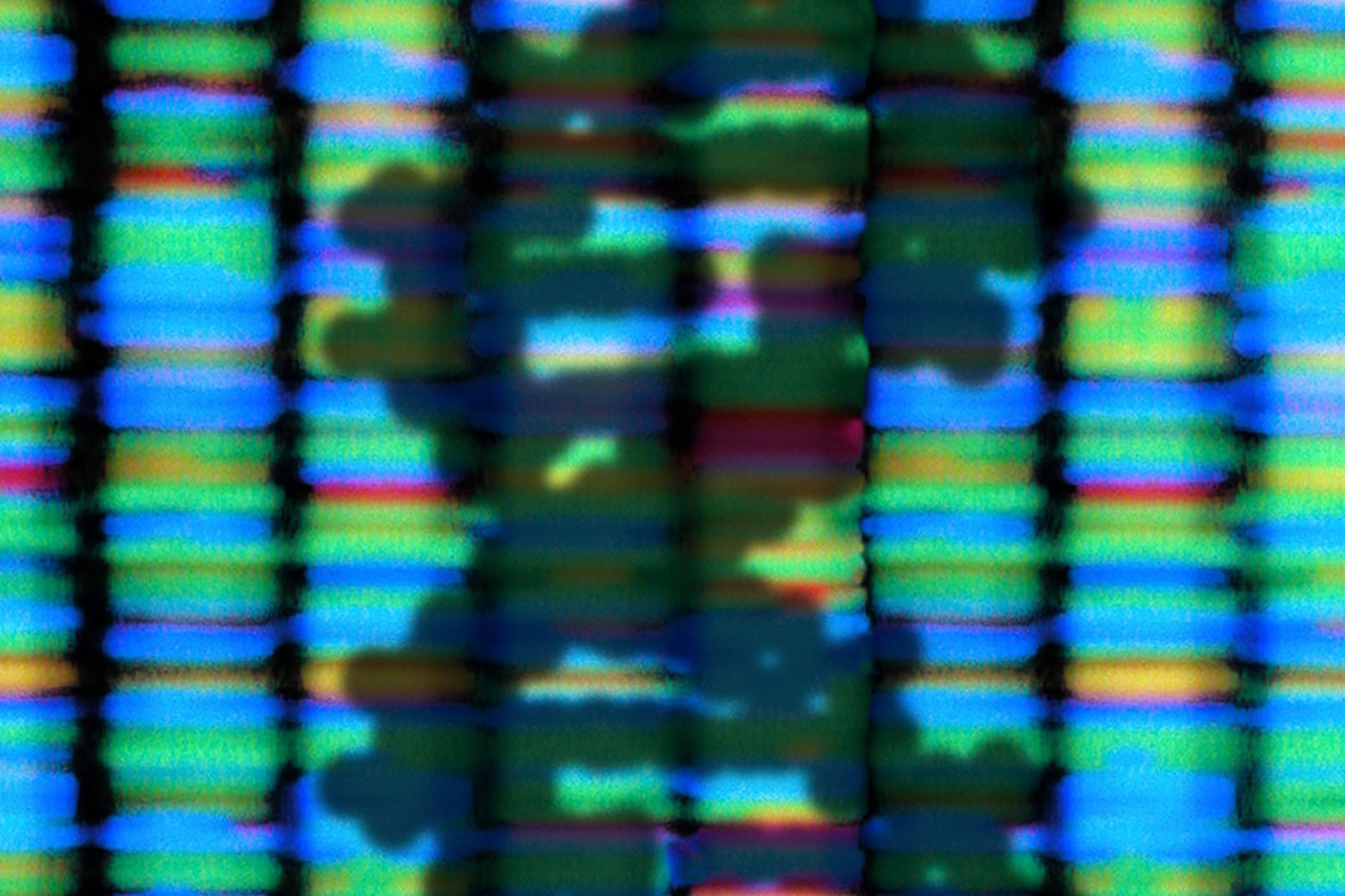
By combining these two different forms of microscopy with computer modelling into a single technique called 3D-CLEM, researchers from the University of Edinburgh have been able to study the structure of human chromosomes in unprecedented detail. Their surprising results showed that, at certain stages in the cell division process, up to 47 percent of our chromosomes are made up of a layer of proteins, called the periphery, which covers the their surface.
The study's lead author, Dr Daniel Booth from the University of Edinburgh, said: 'The imaging technique we have developed to study chromosomes is truly groundbreaking. Defining the structure of all 46 human chromosomes for the first time has forced us to reconsider the idea that they are composed almost exclusively of chromatin, an assumption that has gone largely unchallenged for almost 100 years.'
Although little is known about the periphery, previous work by the University of Edinburgh group has shown that it is needed for a cell to successfully finish the process of cell division and to ensure that the chromosomes do not clump together during division. Based on these observations, it is thought that the periphery may play a role in protecting DNA and that problems with the periphery may contribute to some cancers and birth defects. However, further research will be necessary to confirm this.
Professor William Earnshaw, also from the University of Edinburgh and a co-author of the study, explained: 'We now have to re-think how chromosomes are built and how they segregate when cells divide, since the genetic material is covered by this thick layer of other material.'
This work was carried out in collaboration with the University of Liverpool, UK, the Kazusa DNA Research Institute, Japan and the National Cancer Institute, Maryland, USA, and was published in the journal Molecular Cell.
Sources and References
Related Articles

Light shed on sperm DNA packing process
Crucial role of sperm DNA-packaging protein has been identified in mice, which causes infertility when truncated...
New DNA structure found in human cells
A 'twisted knot' of DNA, known as an i-motif, has been found in living cells for the first time, raising questions about its function...
Loops or spirals? 150-year mystery of what happens to DNA in cell division solved
How DNA is accurately split between cells when they divide has finally been solved by researchers...
Scientists use CRISPR to reveal DNA folding secrets
Scientists have used 'genome surgery' to change the way DNA is packed inside human cells...
3D map of human genome shows DNA 'loops'
A study into the 3D structure of the human genome has revealed the locations of DNA 'loops' at unprecedented resolution...
Leave a Reply
You must be logged in to post a comment.