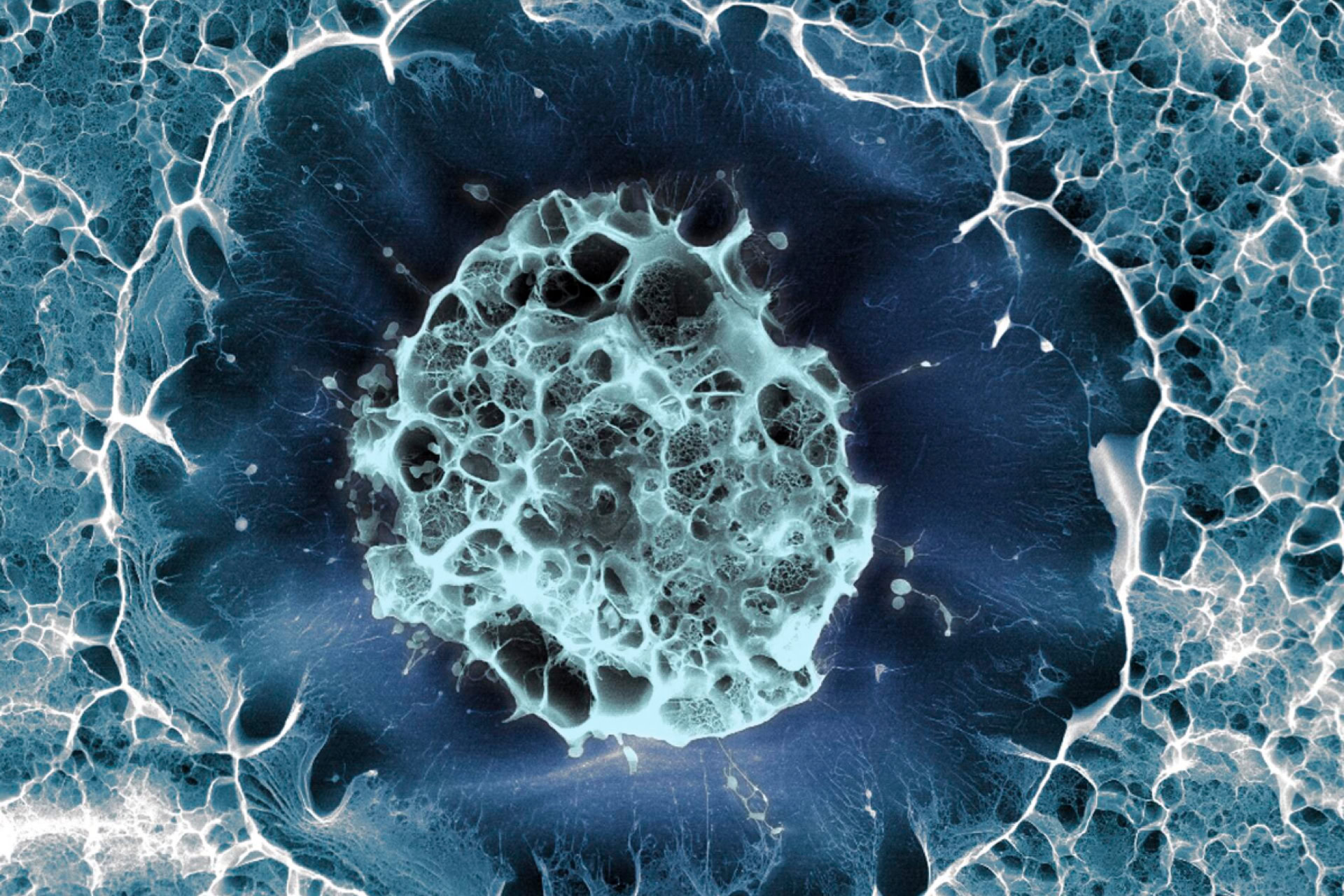
Microscopic nanodevices have been injected inside cells for the first time, allowing researchers to track the beginning of embryo development.
A joint study from the University of Bath and the Instituto de Microelectrónica de Barcelona, Spain has introduced tiny nanodevices inside mammalian cells. This has enabled unprecedented insights into the mechanical forces and internal changes occurring at initial stages of development.
'This is the first glimpse of the physics of any cell on this scale from within,' said senior author Professor Tony Perry. 'It's the first time anyone has seen from the inside how cell material moves around and organises itself.'
Published in Nature Materials, the study developed a silicon-based nanodevice, which along with sperm, was injected into the egg of a mouse. This led to the creation of a healthy, fertilised mouse embryo with an embedded tracking device.
The behaviour of the cell material is hypothesised to be as influential as gene expression to the behaviour and the development of the cell. However, intracellular forces have been poorly understood and difficult to study. Now, these minuscule devices are capable of detecting pulling and pushing forces at very high precision that reveal how the cell material rearranges over time.
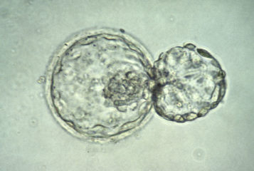
Researchers investigated the mechanical changes occurring inside the cell when a one-cell embryo develops into a two-cell embryo. Measurements were taken by video recordings of the one-cell embryo through a microscope.
The study detected dynamic mechanical forces, ranging from calm periods to forces that were greater than those inside muscle cells. These results also suggest that physical changes inside the cell are programmed from the initial stages of embryonic development.
This new technology could be used in the future to better understand how cells behave under different conditions, tracking the movement of cellular material during ageing and in disease.
Sources and References
Related Articles
Human stem cells grown inside mouse embryos
Scientists have developed a method that dramatically increases the production of human stem cells using mouse embryos...
Development of guidelines for research on stem-cell based embryo models
New international guidelines are being developed to establish ethical parameters for scientists working with human stem-cell based embryo models...
'Living robot' life forms created from frog embryo cells
Scientists at the University of Vermont and Tufts University, Massachusetts have reported building so-called 'biological robots' for the first time...
Leave a Reply
You must be logged in to post a comment.